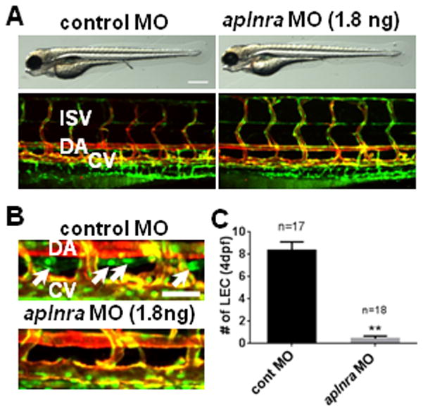
Fig. 1. Lack of aplnra activity causes lymphatic vessel defects.

(A) Gross morphology (top panels) and vascular structures (bottom panels) in 4dpf aplnra MO-injected embryos. (B) The loss of LECs in thoracic duct (white arrows) in 4dpf aplnra MO-injected embryos. (C) Quantification of the number of LECs in control and aplnra MO-injected embryos. LECs within the thoracic duct between 8th and 15th somites were counted. All embryos shown have Tg(fli1a:nEGFP);Tg(kdrl:mCherry) double transgenic background to visualize BECs (shown as yellow) and LECs (shown as green). DA, dorsal aorta; CV, cardinal vein; ISV, inter-segmental vessel. Scale bars are 400μm (A) and 50μm (B).
