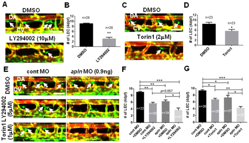
Fig. 4. Apelin and AKT cooperate to promote lymphatic development in zebrafish.

(A) The number of LECs (white arrows) was substantially decreased in embryos treated with a chemical inhibitor of PI3K (LY294002) between 48hpf and 4dpf, therefore, with an attenuated level of AKT activity. (B) Quantification of the number of LECs in 4dpf DMSO or 10μM LY294002 treated embryos. (C) The number of LECs (white arrows) in embryos treated with Torin1 between 48hpf and 4dpf, a chemical antagonist against another upstream regulator of AKT, mTORC1/2, was significantly decreased. (D) Quantification of the number of LECs in 4dpf DMSO or 2μM Torin1 treated embryos. (E) Apelin and AKT may function within the same pathway. Treating embryos injected with a low dose of apln MO with a suboptimal dose of LY294002 (5μM) or Torin1 (1μM) exacerbated the effects of apln MO on the number of LECs (arrows). (F) Quantification of the number of LECs in control or apln MO injected embryos treated with either DMSO or LY294002. (G) Quantification of the number of LECs in control or apln MO injected embryos treated with either DMSO or Torin1. DA, dorsal aorta; CV, cardinal vein. Scale bars are 50μm.
