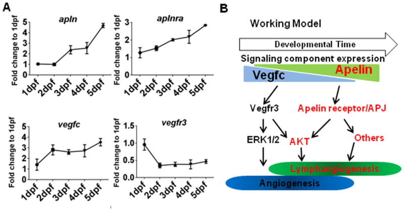
Fig. 6. Apelin and Vegfc signaling is temporally separated for lymphatic vessel formation in zebrafish.

(A) Expression profiles of apln, aplnra, vegfc, and vegfr3 during the first five days of zebrafish development, normalized to actb1. The expression of apln and aplnra gradually increased over time, while the expression of vegfc and vegfr3 is either constant or attenuated. (B) Schematic working model. Apelin and Vegfc signaling appear to have non-overlapping functions during lymphatic development. While functionally redundant in activating AKT, Apelin and Vegfc signaling may be temporally separated and seem to activate specific sets of downstream effectors.
