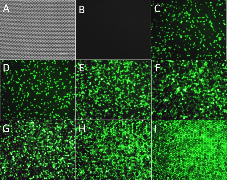
Figure 6.
(a) Typical image of a close-packed single layer droplet array. (b)-(i) Fluorescence images of droplets with different molecule numbers of β-Gal: negative control with no enzyme (b), 0.1 cpd (c), 0.25 cpd (d), 0.5 cpd (e), 0.75 cpd (f), 1 cpd (g), 2 cpd (h), and 3 cpd (i). All the droplets have reacted for 4 h and were single layer close packed. Horizontal scale bar is 100 μm.

