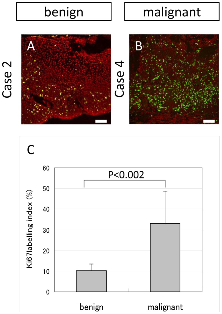
Figure 4. Ki67 expression in OSSN cases.

(A and B) Representative expression pattern of benign cases (A, case 2) and malignant cases (B, case 4). Ki67 positive cells were disseminated both in benign and malignant samples. The expression pattern was comparatively sparse and limited in the basal area in the benign samples (A), while diffuse and dense in malignant samples (B). (C) Ki67 labelling index was significantly (p<0.002) high in the malignant tissue (33.2%) rather than benign tissue (10.9%). Scale bars = 100 µm.
