
Figure 3. Histological structure of ovine vaginal wall.
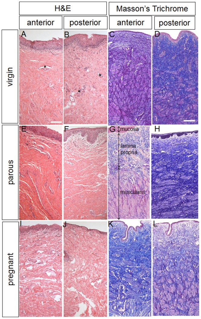
H+E and Masson stained sections showing the anterior and posterior vaginal walls of virgin (A–D), parous (E–H) and pregnant sheep (I–L). * indicates blood vessels. In image E arrows indicate the 3 vaginal layers. Scale bar for H+E sections 100 µm, for Masson sections 250 µm.