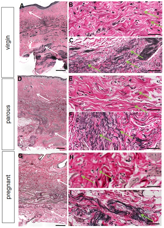
Figure 6. Verhoff's van Gieson staining for elastic fibres (black) on virgin (anterior), parous (posterior) and pregnant (posterior) vaginal tissues.

The white arrows on the full thickness images A, D, G denote the region where the higher magnification images were taken in the lamina propria (B, E, H) and deep muscularis (C, F, I). The green arrows indicate regions of elastic fibres. Scale bars 500 µm (full thickness images) and 50 µm (high magnification images).
