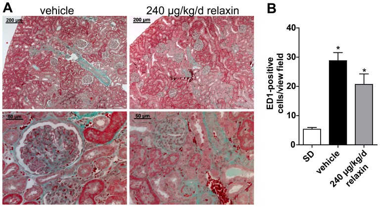
Figure 5. Renal fibrosis and inflammation.
Masson trichrome staining of kidney sections (A) showed increased matrix deposition in vehicle-treated dTGR. Relaxin did not reduce matrix formation in the kidney (representative images). The upper panel shows a higher magnification. (B) Semi-quantification of ED-1-positive cells in the kidney revealed that relaxin did not reduce monocyte/macrophage infiltration in kidney. At least fifteen different areas of each kidney were analyzed. Results are mean ± SEM of 4 animals per group.

