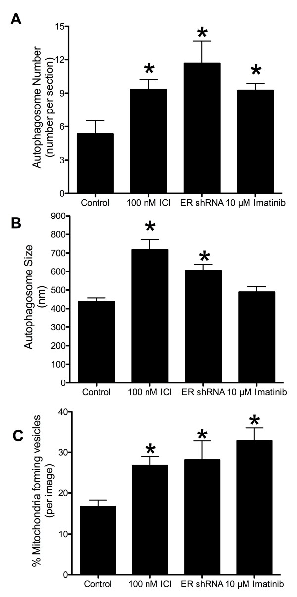
Figure 3.
Autophagosome number, size quantification, and percent mitochondria developing vesicles. Autophagosomes were counted (A) and measured (B) using Image J software from electron microscopy images of LCC9 breast cancer cells. n = 3-5, *p < 0.05. C. Mitochondria were counted and graphed as percent of mitochondria forming vesicles. n = 5-7.

