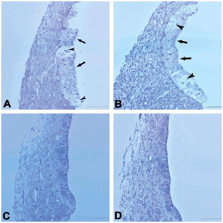
Figure 3.
Cross-sectional views of SV in the basal turn obtained at different times after a single furosemide injection. (A) At 30-min post-furosemide, severe SV edema and breakage were seen, which involved all three cellular layers in some places (short arrows), interrupting SV continuity. MCs were swollen and protruding into the scala media (long arrows), making the surface of the SV appear rough. (B) 90 min after furosemide administration, the SV breakage was not seen but swollen MCs and intermediate cells, large vacuoles across MC (short arrows) and the intermediate and probably basal cell layers remained. Moreover, many MCs still protruded into the SM (long arrows). (C) 12 hr after the furosemide injection, no obvious pathology was seen, compared with the control images. (D) Control. Scale bar: 50 μm.

