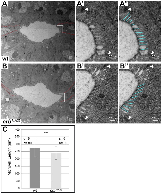
Figure 5. Loss of Crb from the BCs alters the apical membrane structure.
(A–B″) Electron micrographs of cross sections through the large intestines of stage 16 wild-type (A–A″) and homozygous mutant crb11A22 (B–B″) embryos. In A and B, BCs are outlined by red lines, the rectangles indicate areas enlarged in A′, A″, B′ and B″. White arrowheads in A′, A″, B′ and B″ point to the adherens junctions between the BCs and PCs. BCs form longer and more regular microvilli than the PCs in wild-type (A–A″) and homozygous crb11A22 mutant embryos (B–B″). (C) Graph showing the mean length of microvilli in the BCs of stage 16 wild-type and crb11A22 mutant embryos ± standard deviation. s refers to the number of embryos analysed; n refers to the number of microvilli analysed. ***indicate p-value <0.001, assessed by two-sided Student's t-test.

