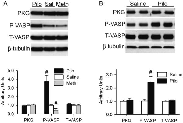
Figure 6. M2 receptor manipulation alters the phosphorylation of a PKG substrate.
Shown are representative images (upper panels) and pooled densitometry data (lower panels) of western blot analyses for PKG, Ser239-phophorylated VASP (P-VASP), and total VASP (T-VASP). β-Tubulin was probed for loading control. Muscarinic receptors in mice (A) were interrogated as described in Figure 1; those in cultured NRVMs (B) are interrogated as described in Figure 5. N= 6 per treatment.

