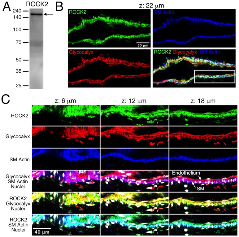
Figure 2. Expression of ROCK 2 in rat mesenteric lymphatic vessels.
A. Western blot for ROCK2. B. Confocal section images of the isolated lymphatic vessel shown in Movie S2. The vessel was labeled to identify ROCK2 (green), glycocalyx that is present on both the endothelial and smooth muscle layers (red), and smooth muscle (SM) actin to distinguish the smooth muscle layer (blue). An overlay of the green, red, and blue channels is also shown. The confocal series included slices taken every 2 μm, and the panel B images correspond to the confocal slice taken 22 μm from the start of the series. The white box in the overlay image indicates the zoom-in area shown in panel C, where images corresponding to confocal slices at 6, 12, and 18 μm through the series are shown. The ROCK2 (green), glycocalyx (red), and smooth muscle actin (blue) channels, along with overlays including nuclei (white) are shown. For the glycocalyx/smooth muscle actin overlay, magenta areas represent overlap of the red and blue labels. This helps distinguish the endothelium, which labels only in red, from the smooth muscle (SM) layer, which is blue/magenta. For the ROCK1/glycocalyx overlay, yellow areas represent overlap. For the ROCK1/SM actin overlay, cyan areas represent overlap. Representative of three experiments.

