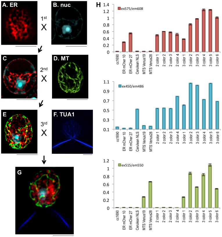
Figure 3. Gene stacking through mating.
An mt+ strain transformed with pBR30 (A) was crossed with an mt- strain transformed with pBR28 (B). Progeny that expressed mCherry in the ER and mCerulean in the nucleus were obtained (C). Cell lines expressing both ER-mCherry and nuclear mCerulean were crossed with cells transformed with mitochondria-targeted Venus (D), to obtain progeny that stably expressed three distinct FPs in three sub-cellular locations (E). These cell lines were crossed to transgenic cells that expressed α-tubulin (TUA1) fused to mTagBFP (F), to obtain progeny that expressed four FPs in four distinct subcellular locations (G). H. Fluorescence plate reader assays of the parents and progeny indicate that FP expression remains stable following matings. Cell lines were assayed for mCherry expression (ex575/em608), mCerulean expression (ex450/em486), and Venus expression (ex515/em550). ER-mCherry parents (ER-mCher 10 and 27), nuclear mCerulean parent (Cerulean NLS) and mitochondrial Venus parents (MTS Venus 19 and 28) are shown along with WT cc1690. 2 color cell lines 1–4 express ER-mCherry and Cerulean-NLS. 3 color cell lines 1–6 express ER-mCherry, nuclear mCerulean and mitochondrial Venus. Scale bars, 5 μm.

