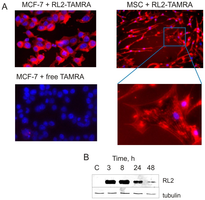
Figure 3. RL2 penetrates into the cells.
A. Fluorescence microscopy analysis of MCF-7 and MSC cells was performed after treatment with RL2-TAMRA conjugate for 1.5 h using the IN CELL Analyzer system. For visualization of nuclei the cells were stained with DAPI. Images are representative of two independent experiments. B. Time-course of RL2 stability in MCF-7 cells. Cells were treated with RL2 (0.2 mg/mL) for the indicated time followed by the preparation of whole-cell lysates, which were then subjected to Western blot with anti-RL2 antibodies. Results shown are representative of five independent experiments.

