
Figure 4. Separation of proteins of RL2-bound fraction.
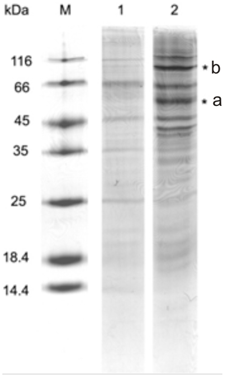
Coomassie stained gel. RL2-bound fractions of MCF-7 lysates were prepared as described in ‘Materials and methods’ section and then were separated by 15% SDS-PAGE gel. Bands indicated (*) were excised followed by in-gel trypsin digestion for mass spectrum analysis. Other bands were not investigated. Shown is representative gel of two independent experiments. Lane 1 – fraction eluted from non-modified Sepharose (control); Lane 2 – fraction eluted from RL2-modified Sepharose with 300 mM NaCl, flow rate was 1 mL/min and detection wavelength was 280 nm. M – molecular weights marker (14.4–116.0 kDa).