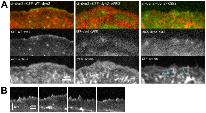
Figure 2. Dynamin2 is targeted to the distal lamellipod via interactions of the dynamin2 stalk and the dynamin2 PRD.
(A) Images from movies of dyn2-depleted U2-OS cells transiently expressing either GFP-WT-dyn2 or GFP-dyn2-ΔPRD, as indicated, and mCh-α-actinin, or mCh-dyn2-K5E5 and GFP-α-actinin, as indicated (see Movie S2); cyan arrowheads highlight GFP-α-actinin-decorated cables induced in cells expressing mCh-dyn2-K5E5. The images of mCh-dyn2-K5E5 and GFP-α-actinin were pseudocolored αs green and red, respectively, to correspond with the other images displayed in this panel. (B) Representative kymographs obtained from pixel-wide lines orthogonal to the leading edge of dynamin2-depleted cells expressing GFP-WT-dyn2 depicting the spatial and temporal distribution of GFP-WT-dyn2. Scale bars: as indicated.

