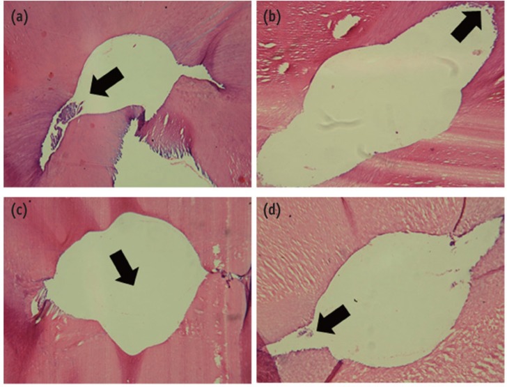
Figure 2.
Histological images from samples of Group 1 (a) and (b) and Group 2 (c) and (d). (a) Remaining pulp tissue in the region of the isthmus (arrow); (b) Debris in the root canal wall (arrow), where the instruments have not touched; (c) Root canal lumen (arrow), which is clean and free of debris; (d) Root canal lumen free of debris, with little remaining pulp tissue in the isthmus area. H. E. stain (×64).

