Fig. 4.
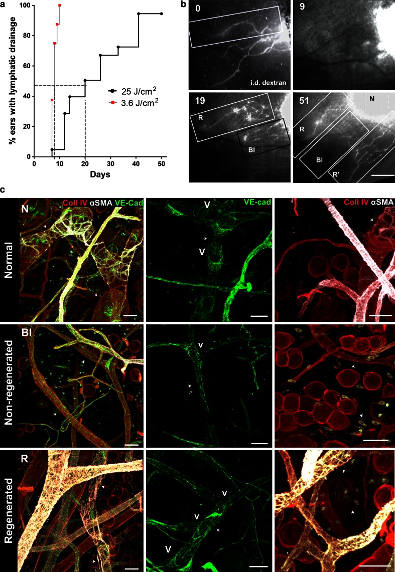
Kinetics of functional recovery and characteristics of regenerated lymphatics after PDT. a Kaplan-Mayer plot showing percent of ears with functional lymphatic drainage versus time for 3.6 versus 25 J/cm2 dose of light, with median recovery times of 8 and 20 days, respectively (p < 0.0001). The regeneration of lymphatic drainage was concluded when at least one collector drained TRITC-dextran injected in the region that was initially injected with verteporfin. b Lymphatic drainage recovery as detected by fluorescence microlymphangiography. Fluorescence microlymphangiography with intradermally (i.d.) injected TRITC-dextran performed on the same ear immediately after PDT (0) and again on days 9, 19, and 51 days after PDT. Three distinct skin zones could be identified: zone with normal (control) (N), blocked and non-regenerated (Bl), and functionally regenerated (arrows) (R). c Representative confocal images of lymphatic vessels in each zone 51 days post-PDT (the same mouse ear as in B and Supplementary Fig. 2) after whole-mount immunostaining for collagen IV (red), α-smooth muscle actin (αSMA, white) and VE-cadherin (green). Left column αSMA-positive pericytes that are normally sparse in control lymphatic vessels (N) are absent in non-regenerated lymphatics (Bl) but overabundant around regenerated lymphatic vessels (R). Middle column Valves (v) that normally exist at the lymphatic junctions (N) were absent at the junctions of non-regenerated lymphatics (Bl) and sparsely located at the junctions of regenerated collectors (R). Right column Myofibroblasts expressing αSMA that are lacking in normal tissue (N) are present in the tissue in areas with non-regenerated (Bl) and regenerated (R) vessels (arrowheads). Arrows indicate lymphatic vessels. Bars in b correspond to: 500 μm, in c to 50 μm

