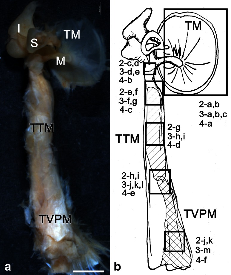
Fig. 1.
Inner side views of the human right TTM ear ossicles (M malleus, I incus, and S stapes), and TM (60-year-old male) (a) The observed sites are shown in b 2-a, b. 3-a-c, 4-a inner surface of the tympanic membrane; 2-c, d, 3-d, e, 4-b neck of the malleus; 2-e, f, 3-f, g, 4-c insertion of the TTM at the malleus; 2-g, 3-h, I, 4-d belly of the TTM, 2-h, i, 3-j-l, 4-e connection region between the TTM and TVPM; 2-j, k, 3-m, 4-f belly of the TVPM; bar 2 mm

