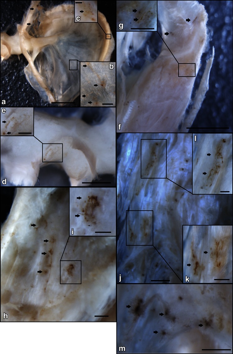
Fig. 3.
The distribution of Calcitonin gene-related peptide-immunoreactive nerve fibers (CGRP-IR NFs) in the inner surface region of the middle ear shown by immunohistochemical staining at the macroscopic level. a CGRP-IR NFs were located in the inner surface of the TM by anti-CGRP-IR NF immunohistochemical reactions (77-year-old female) (bar 2 mm). b Higher magnification of the square in a. CGRP-IR NFs (arrows) were sparsely located on the TM (bar 0.25 mm). c Higher magnification of the square in a. CGRP-IR NFs (arrows) were found in the fibrocartilaginous ring of the pars tensa of the TM (bar 0.1 mm). d Higher magnification of the neck of the malleus (see Fig. 1) (bar 1 mm); e Higher magnification of the square in d. A few CGRP-IR NFs (arrows) were observed (bar 0.5 mm). f The insertion site of the TTM on the malleus (see Fig. 1) (bar 2 mm); g Higher magnification of the square in f. CGRP-IR NFs (arrows) were observed (bar 0.5 mm). h The belly of the TTM (see Fig. 1). CGRP-IR NFs (arrows) were observed on the TTM (bar 0.5 mm). i Higher magnification of the square in h. Mesh-like structures of CGRP-IR NFs (arrows) were observed (bar 0.16 mm). j The connection site between the TTM and the TVPM (see Fig. 1) (bar 1 mm); k Higher magnification of the square in j. CGRP-IR NFs (arrows) were found in the vessel-like structures (bar 0.3 mm). l Higher magnification of the square in j. m The belly of the TVPM (see Fig. 1). Concentrated CGRP-IR NFs (arrows) were observed (bar 0.5 mm)

