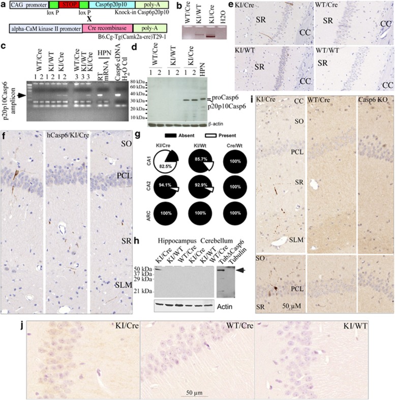
Figure 2.
Transgenic model expresses Casp6 in the CA1 of the mouse hippocampus. (a) Schematic diagram showing the structure of the transgenic hCasp6 cDNA and CaMKIIa-Cre constructs. (b) Ethidium bromide-stained agarose gel showing excision of the Stop sequence in the KI/Cre mouse brain by PCR amplification of the ACL-1 locus (CAG-loxP-STOP-loxP-Casp6p20p10). (c) Ethidium bromide-stained agarose gel showing the hCasp6 amplicon from the KI/Cre but not the KI/WT or WT/Cre mice brain. Numbers represent three different cases. HPN, human primary neurons. (d) Western blotting of human p20p10Casp6 expression in brain of KI/Cre mice. (e) Micrograph of mouse CA1 tissue sections immunopositive for anti-hCasp6 in Casp6 KI/Cre but not in KI/WT, WT/Cre, or WT/WT brains. CC, corpus callosum. Controls to show specificity of the anti-hCasp6 antisera are in Supplementary Figure S2. (f) Immunostained hCasp6 distribution in neurites of the SO, PCL, SR, and SLM of the CA1 region. (g) Percentage of KI/Cre (n=17), KI/WT (n=14), WT/Cre (n=11) CA1, CA2, or ARC immunopositive for hCasp6. Mice were aged from 2 to 20 months. (h) Western blotting showing TubulinΔCasp6 in the KI/Cre hippocampus but not in the cerebellum or in KI/WT and WT/Cre hippocampus and cerebellum. Recombinant full-length tubulin (FL Tub) and Tubulin cleaved by Casp6 (TubΔCasp6) were used as controls. (i) Micrograph showing TubulinΔCasp6 immunoreactivity in similar locations of KI/Cre CA1 as observed for hCasp6. Mice hippocampus shown were from 24-month-old KI/Cre and WT/Cre mice, while the Casp6 KO was 12 months old. (j) Immunostaining of mouse brains with anti-active Casp6 (Biovision)

