
Figure 3.
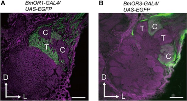
Projection patterns of bombykol- or bombykal-sensitive ORNs to the AL. The axon terminals of bombykol (A) and bombykal (B) receptor neurons were labeled by EGFP using the BmOR1 or BmOR3 promoter-GAL4 lines and a UAS-EGFP line. EGFP was visualized using anti-GFP immunostaining (green). Background staining was carried out using Alexa Fluor 555 (magenta). Representative confocal sections are shown. C, cumulus; T, toroid; D, dorsal; L, lateral. Scale bars: 50 μm. (modified from Sakurai et al., 2011).