Figure 5.
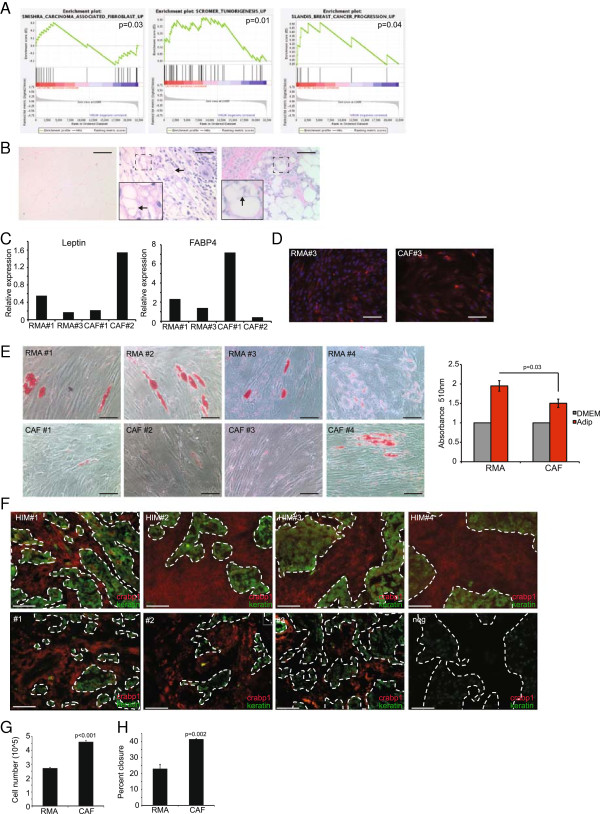
CAFs phenocopy ASC-Ls. (A) Gene sets enriched in ASC-Ls. (B) Representative H & E-stained sections of normal human mammary gland adipocytes (left), human mammary carcinoma (middle) and human xenograft breast cancer (right). Arrows and inset indicate prominent membrane folding. Scale bar = 100 μm. (C) Quantitative RT-PCR of leptin and FABP4 in RMA and CAFs from various patient samples. Data are presented as average 2(-ΔΔCt) ± SEM. (D) Representative image of immunofluorescence of RMAs and CAFs for FABP4 (red) and counterstained for nuclei with DAPI (blue) (n = 3 per group). (E) Representative images of Oil Red O staining and quantification of adipose tissue from reduction mammoplasty (RMA) (n = 8) and carcinoma associated fibroblasts (CAF) (n = 6) treated with adipocyte differentiation media. Data are presented as average absorbance ± SEM. Untreated cell absorbance is set to one. (F) Immunofluorescence of human mammary carcinoma either as xenografts in the HIM model (n = 4) or primary human breast cancer tissues, n = 10. Crapb1 (red) cytokeratin (green) showing both positive (#1 to 3) and negative expression (neg). (G-H) Endothelial cell assays using HUVECs treated with conditioned media collected from RMAs or CAFs, n = 3 per group. Data are presented as means ± SEM. (G) Proliferation assay day 3. (H) Quantification of wound healing assay six hours after addition of conditioned media. Scale bar = 100 μm. ASC-L, lactation-derived adipose stromal cells; crabp1, cellular retinoic acid binding protein-1; DAPI, 4′,6-diamidino-2-phenylindole; HUVEC, human umbilical cord endothelial cells; SEM, standard error of the mean.

