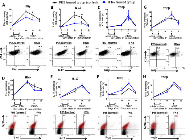
Figure 7.
IFN-α regulates TGF-β and IFN-γ production in macrophages and TGF-β, IL-17, and IFN-γ production in T cells. Draining lymph nodes and spleen cells were collected from mice at days 0, 14, 21, and 28 during the course of AIA and restimulated with mBSA (50 μg/ml) for 24 hours. Brefeldin A (5 μg/ml) and monensin (1 μg/ml) were added 5 hours before FACS analysis. (A-F) Percentage of IFN-γ, IL-17A, and TGF-β expressing lymph node cells of CD4+ cells (A-C) and F4/80+ cells (D, F). (G, H) percentage TGF-β-expressing spleen cells of CD4+ cells (G) and F4/80+ cells (H) during the course of AIA. The data shown are mean values ± SEM of pooled data from two independent experiments (n ≥ 5) with similar results. *P < 0.05; **P < 0.01. Blue line, mice immunized in the presence of IFN-α; black line, control. Below each graph is a representative dot plot of the same cells from mice with or without IFN-α treatment during AIA at day 14 (A-C) and day 21 (D-H). For CD4+ cells, only gated cells are depicted. F4/80+ gated cells are depicted in red, and the figure in the upper right quadrant represents the percentage of cytokine-producing cells among F4/80 (red) cells.

