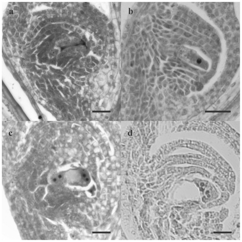
Figure 2. Cytoembryological analysis.
Sexual (a, c) and diplosporous (b, d) development. Bar: 20 μm. (a) Funtional chalazal megaspore and degenerated micropilar megaspores, (b) elongated megaspore mother cell, (c) tetranucleated stage of sexual embryo sac, (d) tetranucleated stage of apomictic embryo sac.

