Figure 2.
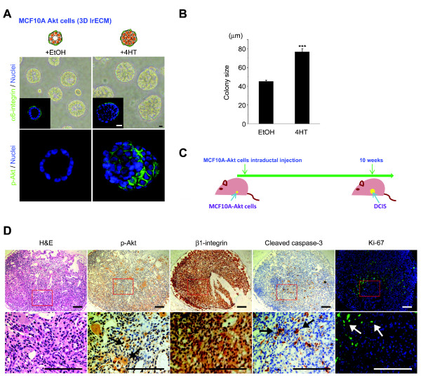
Phosphorylated-Akt up-regulated MCF10A cells form DCIS-like structures in three-dimensional lrECM cultures and in vivo. (A) MCF10A cells form acinar-like structures with hollow lumina when propagated in three-dimensional lrECM. When p-Akt is overexpressed (MCF10A-Akt), the colonies are significantly larger with cells filling the lumina. Phase-contrast micrographs and IF images stained with α6-integrin or p-Akt are shown. Bar = 10 μm. (B) The average colony size is increased in MCF10A-Akt compared to MCF10A. (C) Experimental schema of in vivo study. The MCF10A-Akt cells were injected intraductally into the mouse mammary duct and subsequently generated DCIS-like lesions. (D) H & E, IHC (β1-integrin, p-Akt and cleaved caspase-3) and IF (Ki-67) staining of intraductal xenografts. H & E stained image from the xenograft is almost identical to clinical human DCIS. Bar = 100 μm. DCIS, ductal carcinoma in situ; IF, immunofluorescence; IHC, immunohistochemistry; lrECM, laminin-rich extracellular matrix.

