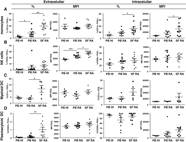
Figure 2.
Characterization of FMS-related tyrosine kinase 3 ligand expression in rheumatoid arthritis paired peripheral blood mononuclear cells (PBMC) and synovial fluid mononuclear cells and in healthy individual PBMC. Intracellular and extracellular expression of FMS-related tyrosine kinase 3 ligand (Flt3L) by all cell types shown in terms of percentage or mean fluorescence intensity (MFI) (cellular marker+Flt3L+). (A) Percentage of extracellular Flt3L by CD14+ monocytes was significantly higher in rheumatoid arthritis (RA) peripheral blood (PB) compared with healthy individuals (HI; n = 10 and n = 5 respectively, P = 0.0400) and in RA synovial fluid (SF) compared with paired PB (n = 10, P = 0.0083). The percentage of intracellular Flt3L by CD14+ monocytes was increased in RA SF compared with paired PB. SF monocytes express higher Flt3L (MFI) compared with RA PB (P = 0.0318 and P = 0.0291 respectively). (B) Expression of extracellular Flt3L (MFI) by CD56+ natural killer (NK) cells in RA SF was significantly higher (P = 0.0191) compared with paired PB. CD56+ NK cells in RA PB expressed significant higher levels of extracellular Flt3L compared with HI peripheral blood mononuclear cells (PBMC; P = 0.0007). (C) CD1c+ myeloid dendritic cells (mDC) in RA SF expressed higher extracellular Flt3L compared with paired RA PB (P = 0.0317). (D) CD304+ plasmacytoid DC in RA SF expressed significant higher (P = 0.0015) levels of extracellular Flt3L compared with paired PB. Bars represent mean (± standard error of the mean) of five to 10 RA patients and HI. *P <0.05, **P <0.01.

