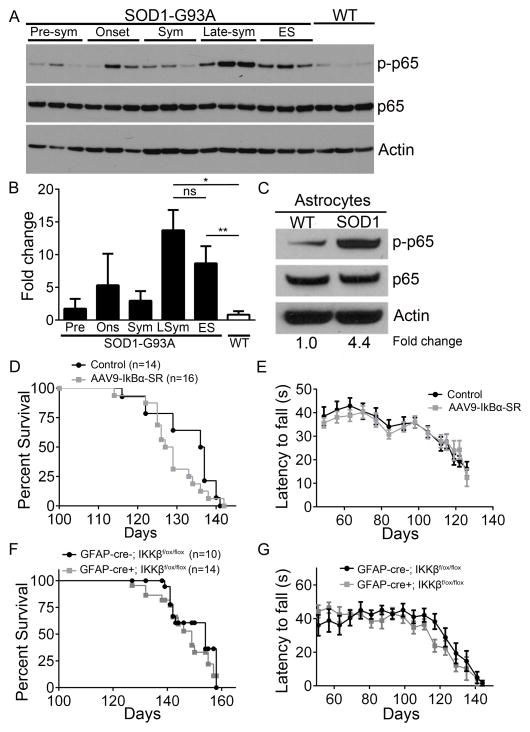
Figure 1. The classical NF-κB pathway is activated with disease progression in the SOD1-G93A mouse model and astroglial NF-κB inhibition does not confer neuroprotection.
(A and B) Immunoblot (A) and fold changes (B) of lumbar spinal cord protein isolated from WT mice at 120 days and from SOD1-G93A female mice at pre-symptomatic (Pre), onset (Ons), symptomatic (Sym), late-symptomatic (LSym), and end-stage (ES). phospho-p65 (top) total p65 (middle) and Actin (bottom). n=3. Additional data shown in Supplemental Figure S1.
(C) Immunoblot of protein isolated from astrocytes obtained from late-sym SOD1-G93A and WT littermates.
(D and E) Kaplan-Meier survival curve (D) of SOD1-G93A mice injected with AAV9-IκBα-SR (median survival=128, n=16) and non-injected controls (median survival=136.5, n=14) and motor performance on accelerating rotarod test (E).
(F and G) Kaplan-Meier survival curve (F) of SOD1-G93A; IKKβf/f; GFAP-cre- (median survival=154, n=10) and SOD1-G93A; IKKβf/f; GFAP-cre+ mice (median survival=149, n=14) and motor performance on accelerating rotarod test (G).
All band intensities were normalized to p65/Actin. Error bars represent s.e.m. *, P<0.05, **, P<0.01. See also Figure S1 and S2.

