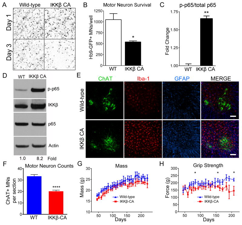
Figure 7. NF-κB activation in wild-type microglia induces MN death.
(A and B) Representative microscopic fields (A) and counts (B) of Hb9-GFP+ MNs after 1 day (24 hours) and 3 days (72 hours) in co-culture with WT microglia (white bar) or WT microglia with constitutively active IKKβ (IKKβCA) (black bar).
(C) Quantification of NF-κB activation (phospho-p65), normalized to total p65, both determined by ELISA.
(D) Immunoblot of lumbar spinal cord protein from WT and IKKβCA mice. The blot was probed for p-p65 (top), IKKβ (top middle), p65 (bottom middle) and Actin (bottom). p-p65 band intensities normalized to p65/Actin. Fold change determined by densitometry in Image J.
(E) Immunohistochemistry of lumbar spinal cords of WT and IKKβCA littermates at 8 months. Iba1-positive microglia (red), GFAP-positive astrocytes (blue), ChAT-positive MNs (green). Scale bar = 100 microns
(F) Counts of ChAT+ MNs in ventral horn of lumbar spinal cord from 8-month old IKKβCA and WT littermates. (n=3)
(G and H) Mass (G) and grip strength (H) of IKKβCA (n=6) and WT littermates (n=8).
Error bars represent s.e.m. *, P<0.05, **, P<0.01, ****, P< 0.0001. See also Movie S6, Figure S5 and Figure S6.

