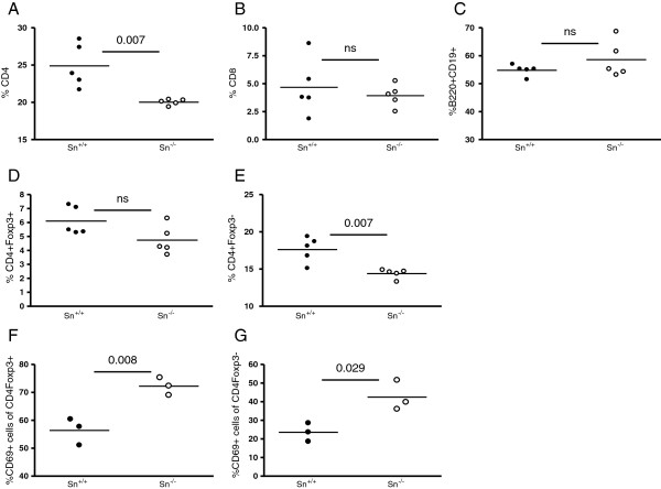
Figure 5.
Flow cytometry of splenocytes from diseased sialoadhesin (Sn)+/+ and Sn−/− New Zealand black x New Zealand white F1 (NZBWF1) mice. (A-G) Splenocytes were isolated from 28-week-old diseased Sn+/+ (filled circles) and Sn−/− (open circles) NZBWF1 mice. Each circle represents an individual mouse. Statistical analysis was done using the Mann–Whitney test and P-values are indicated; ns, not significant.

