
Figure 4.
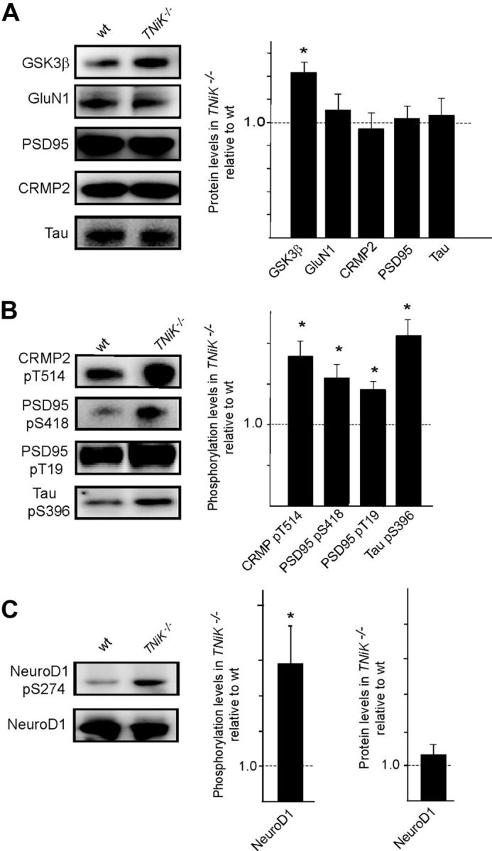
Elevated GSK3β levels in TNiK−/− mice. A, Immunoblots of GSK3β, GluN1, PSD-95, CRMP2, and Tau in PSD lysates from wt and TNiK−/− mice. Histograms show protein levels in TNiK−/− mice PSD normalized to wt levels. B, PSD lysates were immunoblotted with phospho-specific antibodies showing increased phosphorylation of CRMP2 threonine 514, PSD-95 serine 418 and threonine 19, and tau serine 396 in TNiK−/− mice. C, Hippocampal lysates were immunoblotted with phospho-specific antibodies to serine 294 on NeuroD1 (top) and total NeuroD1 (bottom) and levels in TNiK−/− mice quantified relative to wt in histograms. Images show representative Western blot assays from at least triplicate experiments. Asterisk indicates significant difference to wt (p < 0.05).