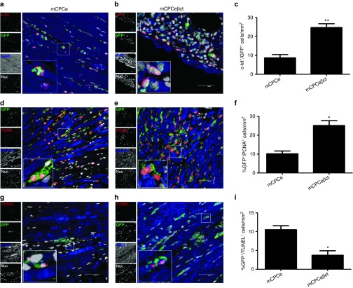
Figure 4.
CPC survival and proliferation is enhanced with βARKct engineering in hearts after acute myocardial infarction. (a–b) Increased c-kit+/GFP+ cells within hearts transplanted with hCPCeβct compared with hCPCe transplanted hearts after 3 days of infarction stained with GFP (green) c-kit (red), sarcomeric actin (blue), and nuclei (white) Scale bar = 40 µm. (c) Quantitation of c-kit+/GFP+ cells in hCPCeβct and hCPCe transplanted hearts. (d–f) Increased PCNA+/GFP+ cells in hCPCeβct hearts compared with hCPCe stained with PCNA (red), GFP (green), sarcomeric actin (blue), and nuclei (white) along with corresponding quantitation. (g–i) Reduced TUNEL+/GFP+ in hCPCeβct hearts compared with hCPCe stained with TUNEL (red), GFP (green), sarcomeric actin (blue), and nuclei (white) along with corresponding quantitation. hCPCe vs. hCPCeβct *P < 0.05, **P < 0.01, ***P < 0.001. GFP, green fluorescent protein; hCPC, human cardiac progenitor cell.

