Figure 1.
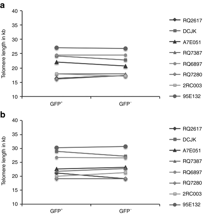
Comparison of telomere lengths in transduced and untransduced blood cells. Quantitative PCR for mean telomere length was performed on genomic DNA from peripheral blood (a) GFP+ or GFP− granulocytes and (b) GFP+ or GFP− lymphocytes from eight rhesus macaques transplanted with lentivirally transduced autologous CD34+ cells 1–13.5 years before. The panels show the telomere length in kilobases (kb) on the y axis for GFP+ and GFP− (a) granulocytes or (b) lymphocytes from each animal. The time of sample collection after transplantation for each animal is given in Table 1. Each sample was run in triplicate, and the mean values are shown as individual points on the plots. There was no significant change in telomere length, calculated as the mean of the triplicate averages for changes in telomere length, between the GFP+ and GFP− cells in either the granulocyte (P = 0.1336) or lymphocyte (P = 0.5183) populations, via a paired Student's t-test.

