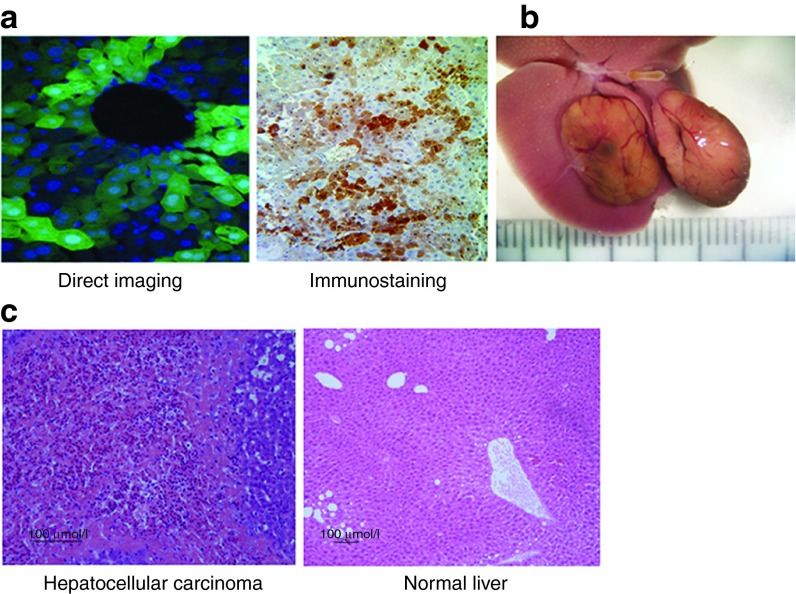
Figure 1.
Transgene expression in hepatocytes and hepatocellular carcinoma development following transduction. Fetal mice were transduced with FIV-hAAT-EGFP at ~40 transducing units per cell. (a) Liver biopsies were analyzed for EGFP expression 5 months posttransduction by direct visualization (left panel; magnification 400×) and immunostaining (right panel; magnification 200×). (b) A representative photo image of two liver tumors that developed in one of the three mice ~1 year posttransduction. The scale on the ruler is 1 mm. (c) H&E staining of tissue sections taken from a liver tumor that developed in mouse M1 (left panel) and from the surrounding liver tissue (right panel). FIV, feline immunodeficiency virus; hAAT, human α-antitrypsin.

