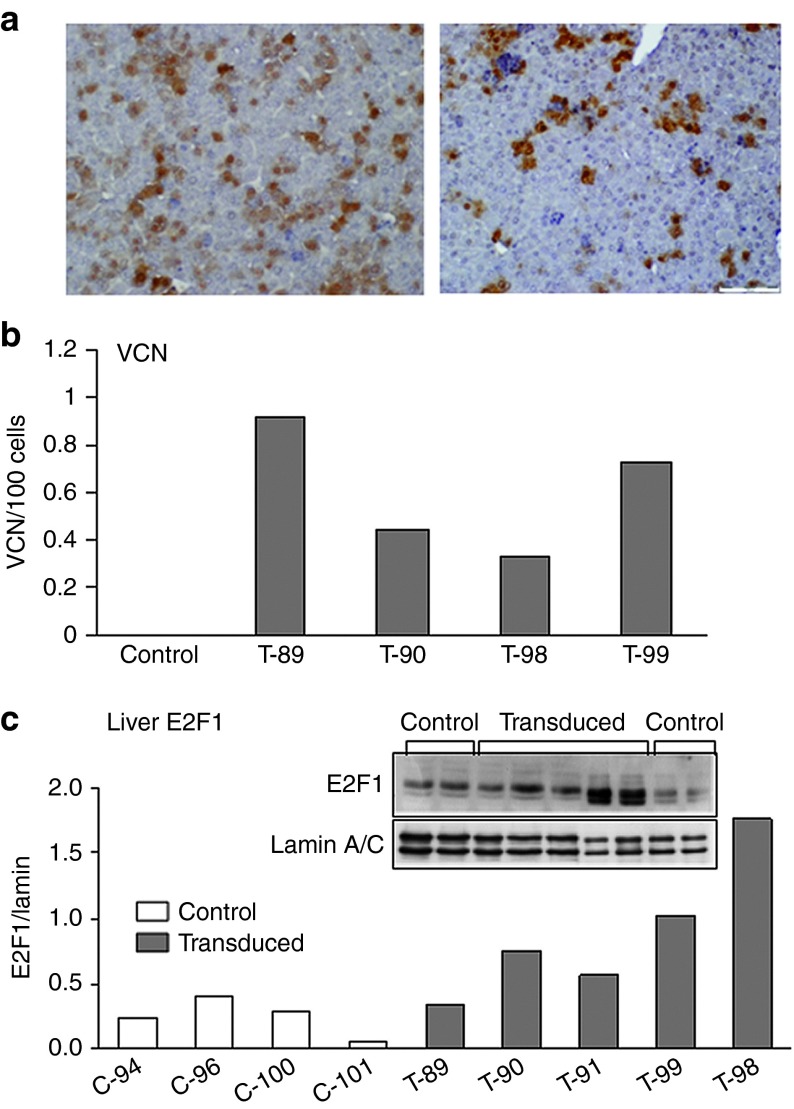
Figure 3.
Analysis of livers from 10-day-old mice after in utero transduction. Fetal mice (at 16 days gestation) were transduced with FIV-hAAT-EGFP lentiviral vector at MOI ~40 and the livers were harvested 2 weeks posttransduction. (a) Tissue sections from livers of transduced mice were analyzed for EGFP expression by immunostaining. Left panel: 36% EGFP-positive cells; right panel: 15% EGFP-positive cells. (b) Vector copy number (VCN) per 100 cells was determined by quantitative polymerase chain reaction on genomic DNA extracted from transduced (T) and untransduced (control) siblings. (c) Nuclear protein levels of E2F1 were determined by immunoblotting analysis of livers of five transduced mice (T) and four untransduced siblings (C). Anti-lamin A/C was used as a loading control. Each column represents one mouse. FIV, feline immunodeficiency virus; hAAT, human α-antitrypsin.

