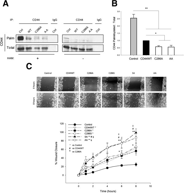
Figure 3.
Reduced CD44 palmitoylation is paralleled by increased cell migration. (A) Biotin-1-biotinamido-4-(4′-(maleimidomethyl)cyclohexanecarboxamido)butane (biotin-BMCC) assays were used to measure palmitoylated CD44 (CD44-Palm) in cells expressing representative single and double palmitoylation mutants, and were compared with total CD44 levels (CD44-Total). Isotype-matched IgG was used as a negative control for CD44 immunoprecipitations (IPs), and omission of hydroxylamine (HAM) reagent was a BMCC-negative control. CD44-Palm was reduced in cells overexpressing CD44 palmitoylation-impaired mutant constructs. (B) Densitometric quantification of multiple experiments confirmed significant reductions in CD44-Palm relative to CD44-Total in mutant cells. Error bars, standard error of the mean (SEM); n = 3. *P < 0.05; **P < 0.01, Student’s t test. (C) Phase contrast micrographs of scratch-wound migration assays performed in MDA-MB-231 cells transfected for 48 hours with CD44WT or CD44 single-site (C286A, C286S) or double-site (SA, AA) palmitoylation-impaired mutants. The graph was constructed by expressing wound width measurements at each time point relative to its cognate time = 0 value. Cell migration was significantly enhanced in mutant-expressing cells compared with control cells, as indicated in the graphical representation of multiple experiments. Error bars, SEM; n = 3. #φ*P < 0.05; **P < 0.01, two-way analysis of variance. WT, wild-type.

