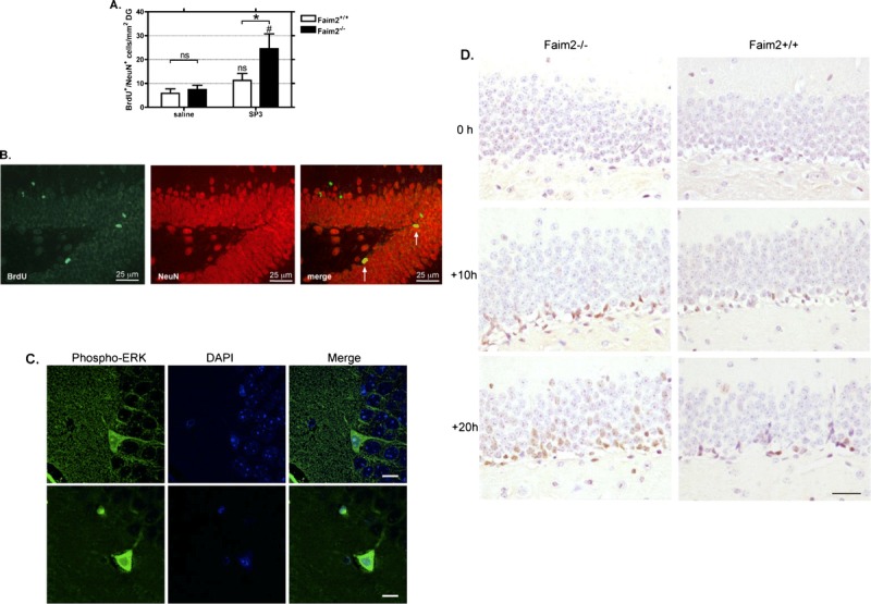
FIGURE 6.

Fas apoptotic inhibitory molecule 2 (Faim2)–dependent hippocampal neurogenesis after Streptococcus pneumoniae type 3 (SP3) meningitis. (A) Quantification of the density of BrdU-immunopositive/NeuN-immunopositive cells in the dentate gyrus reveals a significant increase of newly formed differentiated granule neurons in Faim2−/− mutants versus littermates at 13 weeks after infection indicating increased neurogenesis in knockout mice after meningitis (mean ± SEM; *, p < 0.05 Faim2+/+ [SP3] vs Faim2−/− [SP3]; #, p < 0.01 Faim2−/− [saline] vs Faim2−/− [SP3]). (B) Detection of BrdU (green) and NeuN (red) by double-label fluorescence immunohistochemistry. Merge of both markers reveals newly formed neurons 13 weeks after bacterial meningitis in a Faim2−/− mouse. (C) Confocal images of sections from infected Faim2+/+ mice stained with phospho-ERK1/2 (green) and DAPI (blue) (scale bar = 10 μm). (D) Staining samples of phospho-ERK1/2 in the DG at 0, 10, and 20 hours after infection in Faim2−/− and Faim2+/+ mice showing a time-dependent increase and a tendency for a more pronounced ERK immunoreactivity in Faim2−/− mice versus Faim2+/+ mice.
