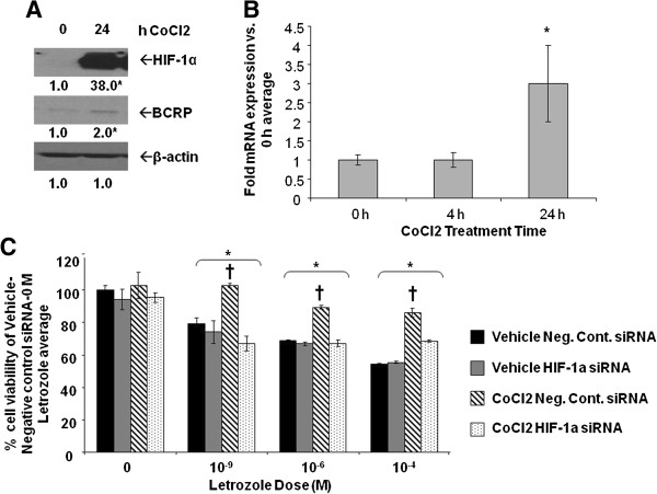
Figure 10.

Effect of CoCl2on MCF-7Ca protein expression and cell viability. A-B, MCF-7Ca cells were incubated in steroid-free media and then treated with 100 μM CoCl2 for 0 to 24 hours. A) Total protein was extracted and HIF-1α, BCRP, and β-actin were analyzed by Western blot analysis. Shown are representative blots and overall densitometry results of n = 6 independent cell samples/group. Densitometry results are expressed as mean fold-change in protein levels compared to 0 hours after normalization to β-actin (mean ± SD of n = 4 independent cell samples/group, *versus 0 hours; P = 0.0005 for HIF-1a; P = 0.0065 for BCRP, two-sided t-test). B) Total RNA was extracted and BCRP mRNA and 18S rRNA were analyzed by real-time RT-PCR analysis. Results are expressed as the mean fold-change in mRNA levels compared with 0 hours after normalization to 18S rRNA (mean ± SD, n = 4 independent cell samples/group; *versus 0 hours, P <0.001; overall P = 0.0002, one-way ANOVA). C) Viability of cells was measured by MTT assay after five days of treatment with increasing doses of letrozole following 48 hours pre-treatment with or without 100 μM CoCl2 and HIF-1α siRNA. Results are expressed as mean percent of 0 μM letrozole-without CoCl2-with negative control siRNA (mean ± SD of n = 6 independent samples/group; *versus vehicle-negative control siRNA-0 μM letrozole, P <0.001; † versus vehicle-HIF-1α siRNA and CoCl2-HIF-1α siRNA; effect of letrozole P <0.0001, effect of pre-treatment (vehicle/CoCl2 and negative control siRNA/HIF-1α siRNA) P <0.0001, interaction between letrozole dose and pre-treatment P <0.0001; two-way ANOVA). ANOVA, analysis of variance; BCRP, breast cancer resistant protein; HIF-1α, hypoxia inducible factor 1 α subunit; MTT, 3-[4,5-dimethylthiazol-2-yl]-2,5 diphenyl tetrazolium bromide; n, number; SD, standard deviation.
