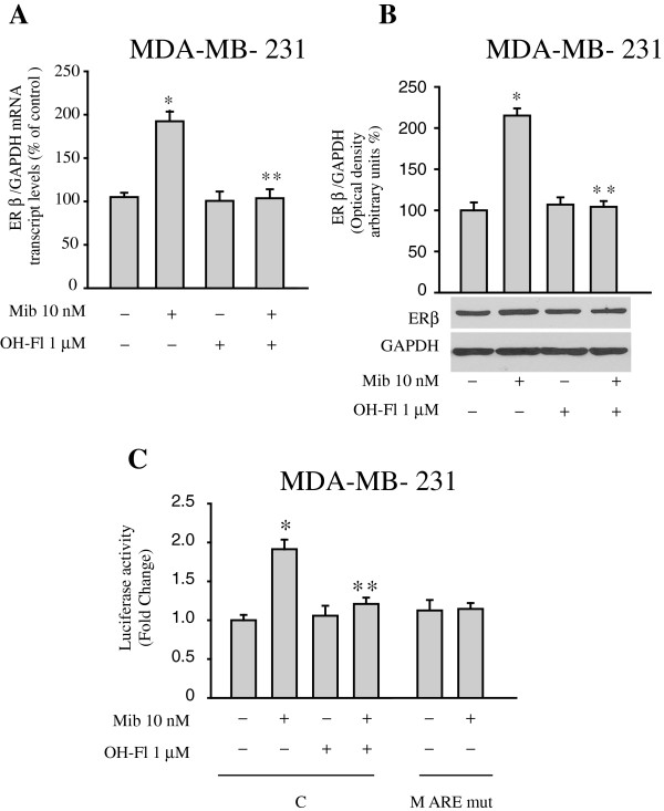
Figure 5.
Mibolerone increases ER beta expression in MDA-MB-231 breast cancer cells. (A) Real-time PCR for ER beta mRNA expression in cells treated with vehicle (−) or mibolerone (Mib) 10 nM in the presence or absence of OH-Fl for 48 hours. Each sample was normalized for its GAPDH mRNA content. Data represent the mean ± S.D. of values from three separate RNA samples expressed as the percentage of the control assumed to be 100%. (B) Bottom panel, Western blot analysis of ER beta in total protein extracts from cells treated with vehicle (−) or Mib 10 nM in the presence or absence of OH-Fl for 48 hours. GAPDH was used as loading control. Upper panel, the histograms represent the mean ± S.D. of three independent experiments in which band intensities were evaluated in terms of optical density arbitrary units and expressed as the percentage of the control assumed to be 100%. (C) ER beta luciferase promoter activity in cells transfected and treated as indicated. The values represent the means ± S.D. of three different experiments each performed in triplicate. *, P <0.05 compared to vehicle-treated cells. **, P <0.05 compared to Mib-treated cells. ER, estrogen receptor.

