Figure 6.
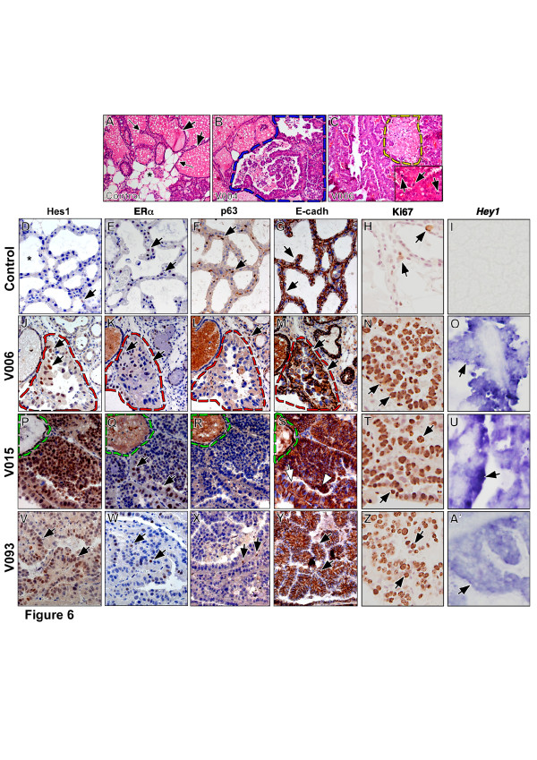
MMTV-Cre; N1ICD transgenic mice develop papillary tumors. (A-C) H&E stainings; (D-H, J-N, P-T, V-Z) immunostainings; (I, O, U, A') in situ hybridization. In (D-G, J-M, P-S,V-Y), sections are counterstained with toluidine blue; in (H, N, T, Z) sections are counterstained with Mayer's Hematoxylin. (A) Normal lactating breast. Greatly expanded secretory lobules (arrows) containing milky secretion are surrounded by breast epithelium (small arrows). Adipose tissue containing lipid vesicles (asterisk) is also abundant. (B) Transgenic female V004. Tumor tissue coexisting with normal tissue is demarcated by a blue dashed line. Breast architecture is widely disorganized. (C) Transgenic female V006. Large necrotic areas (yellow dash line) are observed. Inset shows mitosis (arrows) in tumor tissue. (D) Hes1 expression (brown nuclei, arrow) is rare in normal breast epithelium. Asterisk indicates milky secretion. (E, F) estrogen receptor (brown nuclei, arrows in E) and p63 expression (brown nuclei, arrows in F) in normal breast epithelium. (G) E-cadherin expression in the membrane of breast epithelial cells. (H) Few signs of proliferation in normal lactating breast as indicated by Ki67 staining in only a few cells (brown nuclei, arrows). (I) Normal lactating breasts show no Hey1 expression. (J-O) V006 transgenic female. Tumor area is demarcated by a red dashed line. Note the increased Hes1 expression (J, brown nuclei, arrows). (K) estrogen receptor expression (arrows). (L) p63 expression is observed in the non-pathological tissue (arrow) surrounding the tumor. (M) E-cadherin is expressed in membrane of epithelium. (N) Proliferation can be observed throughout the tumor by Ki67 staining (brown nuclei, arrows). (O) Expression of Hey1 in tumor tissue (arrow). (P-U) V015 transgenic female. Normal tissue is demarcated by green dashed line. (P) Hes1 expression is strongly up-regulated in tumor cells. (Q) Estrogen receptor (arrows) expression is more widespread. (R) p63 expression is very reduced in tumor tissue. (S) E-cadherin is expressed in membrane (arrow) but also in cytoplasm (arrowheads) of tumor cells. (T) Strong proliferation shown by Ki67 staining (arrows). (U) Hey1 expression in tumor (arrows). (V-A') V093 transgenic female. (V) Widespread Hes1 expression in tumor (brown nuclei, arrows). Estrogen receptor (W, arrows) and p63 expression (X, arrows) in tumor. (Y) E-cadherin expression in tumor (arrows). (Z) Strong proliferation in tumor shown by Ki67 staining (arrows). (A') Expression of Hey1 in tumor (arrow).

