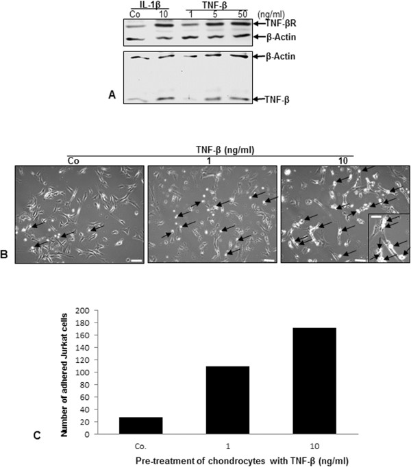
Figure 7.
Upregulation of TNF-β and TNF-β receptor expression in chondrocytes and adhesiveness of T lymphocytes to chondrocytes by TNF-β. (A) Serum-starved chondrocytes were grown to subconfluence in monolayer culture and either left untreated (Co) or treated with 10 ng/ml interleukin 1β (IL-1β) or with 1, 5 or 50 ng/ml tumor necrosis factor β (TNF-β) for 12 hours. Whole-cell lysates were prepared, and samples were examined by Western blot analysis with antibodies against TNF-β and TNF-β receptor (TNF-βR). Western blots shown are representative of three independent experiments. The housekeeping protein β-actin served as a loading control. (B) Chondrocytes were grown to subconfluence in monolayer culture (spindle-shaped to elongated cells) and were either left untreated (Co) or treated with 1 or 10 ng/ml TNF-β for 12 hours, washed and cocultured with Jurkat cells (round to ovoid cells) for 4 hours. After being washed with phosphate-buffered saline, adhesion of T lymphocytes (arrows) to chondrocytes was evaluated by light microscopy. Original magnification, ×100; bar, 30 nm. Inset: original magnification, ×400; bar, 3 nm. (C) The number of Jurkat cells adherent to chondrocytes was estimated and quantified by counting ten microscopic fields per culture. Chondrocytes were either left unstimulated (Co) or stimulated with 1 or 10 ng/ml TNF-β for 12 hours.

