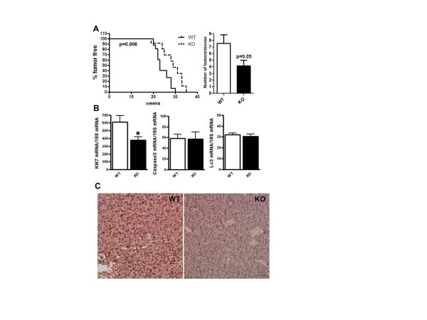
Figure 1.
Tumor onset, multiplicity and cell proliferation were suppressed in COX-2MECKO tumors. COX-2MECKO tumors are denoted as KO. (A) Percent of tumor free mice against weeks of age. Mean tumor free time for COX-2MECKO mice was 29 weeks versus 23 weeks for WT (left graph, n = 13 to 19). The right graph shows tumor multiplicity as number of tumors per mice at necroscopy (n = 14 to 18). (B) Gene expression levels of Ki67 (proliferation), Caspase3 (apoptosis) and Lc3 (autophagy) in whole tumors by Q-PCR (n = 8 to 18). (C) Immunohistochemistry staining for Ki67 (dark red-brown) in sections of paraffin embedded WT and COX-2MECKO tumors (image shown is representative of n = 4). Cell nuclei are counterstained with hematoxylin. The bar on the KO panel indicates 20× magnification. Data in column graphs are mean ± sem. P values are compared to WT; *P < 0.05. COX, cyclooxygenase; KO, knock out; WT, wild type.

