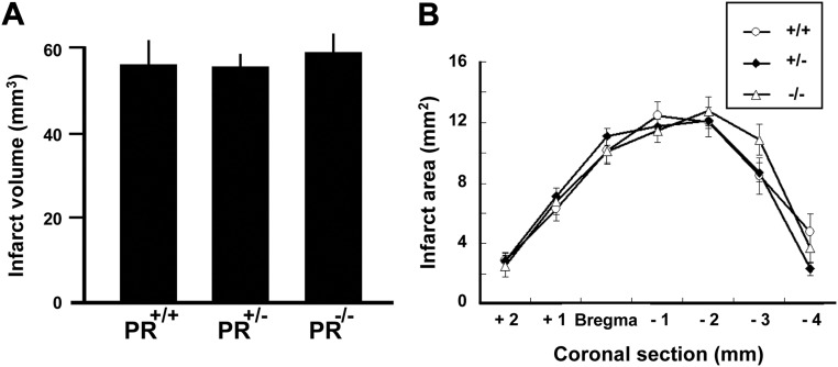
Fig. 4.
Endpoint measures at 48 h after MCAO. A, Total brain infarct volume in PR+/+, PR+/−, and PR−/− mice. There were no significant differences between the three groups. There was also no effect of the PR genotype on lesion volume in cerebral cortex and subcortical structures (not shown). B, The areas of damaged brain tissue measured on seven successive 1-mm-thick brain sections (section 3 = bregma level) were similar in PR+/+, PR+/−, and PR−/− mice. Data represent means ± sem; n = 15 per group.

