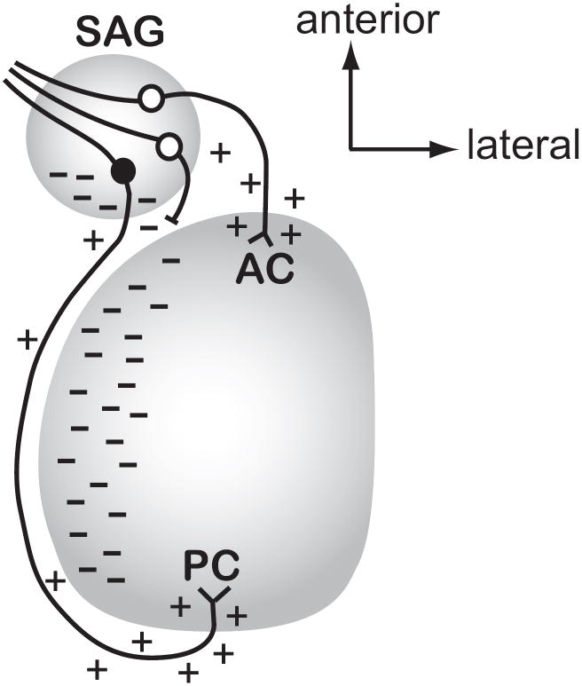
Figure 6. Model for how Slit activity may direct afferent outgrowth into the otocyst in vivo.
This figure predicts where Slit1 and/or Slit2 proteins (indicated as dashes) are localized in the normal ear at the time when afferents to the anterior and posterior crista are projecting from the SAG towards their targets (approximately HH17-22). This model takes into account the experimental observation that afferents projecting to the anterior vs. posterior cristae demonstrate differential responsiveness to Slits as a mechanism by which these two populations of axons are directed to grow in opposite directions. We predict that the diffusion of Slit proteins (indicated by dashes) from the posterior SAG and medial otic vesicle will repel anterior crista afferents (white cell bodies) from initiating trajectories in the posterior direction. If they do project posteriorly, they would be prevented from advancing (shown as an axon with a blocked process) and may then be redirected anteriorly to reach their target. In contrast, a neuron seeking to innervate the posterior crista (black cell body) is insensitive to the same Slit cues, allowing it to pass through or alongside territories that have an abundance of Slit proteins. The model also depicts putative attractive cues (shown as + symbols) along the pathways or emanating from the targets, although additional unknown repulsive cues (not shown) may also confine axonal trajectories to specific pathways. Abbreviations: AC, anterior crista; PC, posterior crista; SAG, statoacoustic ganglion.

