Fig. 1.
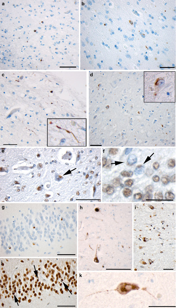
TDP-43 proteinopathy in select positive cases and positive controls. a–b Immunoreactivity to p-TDP-43 in the medial temporal lobe of an 89-year-old female who died 7 years following a single moderate/severe TBI (scale bar 100 µm). c Immunoreactivity to p-TDP-43 in the medial temporal lobe of a 76-year-old female control who died acutely following a myocardial infarction and had no previous history of dementia or other neurological disease (scale bar 100 µm). d p-TDP-43 immunoreactivity in the CA1 region of hippocampus of an 18-year-old male who died 10 h following severe TBI (scale bar 100 µm). e, f Nuclear clearing of TDP-43 in cells following TBI as evidenced using the pi-TDP-43 antibody (same case as a, b) scale bar e100 µm, f 30 µm. g, j Granule cells of the dentate gyrus showing TDP-43 proteinopathy in a case of dementia pugilistica demonstrated using antibodies reactive to p-TDP-43 (g) and pi-TDP-43 (j) (scale bar 70 µm). i Extensive TDP-43 pathology observed in the collateral sulcus of the same case of dementia pugilistica (scale bar 100 µm). h A brainstem neuron reactive for the pi-TDP-43 antibody in a case of ALS. Note the clearing of the nucleus (scale bar 50 µm). k Similar cytoplasmic accumulation with nuclear clearing is observed in a case of FTLD (scale bar 50 µm)

