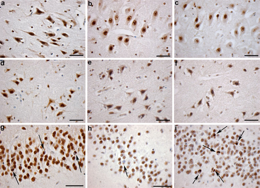
Fig. 3.
Immunoreactivity to full-length pi-TDP-43, and the N-terminus and C-terminus of TDP-43. Cytoplasmic and nuclear immunoreactivity specific for a pi-TDP-43, b the extreme N-terminus of TDP-43 and c the extreme C-terminus of TDP-43 in a 41-year-old male who died 20 h following TBI caused by a fall. Scale bars a–c 50 µm. d–f Cytoplasmic and nuclear immunoreactivity specific for d pi-TDP-43, e the extreme N-terminus of TDP-43 and f the extreme C-terminus of TDP-43 following TBI in a 48-year-old male who died 3 years following injury caused by a fall. Scale bars d–f 50 µm). g Cytoplasmic TDP-43 inclusions with associated nuclear clearing identified using pi-TDP-43 specific antibody in the granule cells of the dentate gyrus in a case of dementia pugilistica (pathology indicated by arrows). h Cytoplasmic TDP-43 inclusions with associated nuclear clearing identified using an antibody specific for the extreme N-terminus of TDP-43 in the same case and region as g. Pathology indicated by arrows. i Cytoplasmic TDP-43 inclusions with associated nuclear clearing identified using an antibody specific for the extreme C-terminus of TDP-43 in the same case and region as g, h. Pathology indicated by arrows. Scale bars g, h 70 µm)

