
Figure 3. Immunocytochemical Staining of Axons Following Dynamic Stretch Injury.
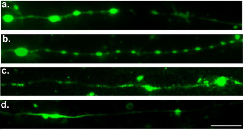
Immunofluorescent images of injured axons (3hr post-injury) displaying a series of swellings along the axonal length like beads on a string. Swellings display accumulations of (a) tubulin, (b) the microtubule-associated protein tau, (c) amyloid precursor protein (APP), and (d) neurofilament (NF200). Scale bar, 10 μm.