
Figure 4. Microtubule Breakage and Loss in Varicose Axonal Swellings 3 Hours After Dynamic Stretch Injury of Axons.
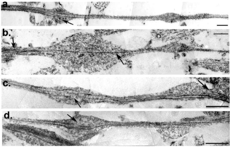
(a-d) TEM images of axons at 3hr post-injury were stitched together to reconstruct a panoramic image of an axon segment. Individual microtubules display selective breakage within axon swellings (a,c,d arrows). While breakage or loss of microtubules can be seen in association with swelling, other microtubules can be observed traversing the swollen region intact. Specifically, (b) shows a solitary intact microtubule traversing a large swelling (arrow) whereas multiple microtubules can be observed in the adjacent non-swollen region from which it emerged (arrow). This sole remaining microtubule feeds into a subsequent but separate swelling. Scale bars: 500 nm.