
Figure 1. Immunohistochemical findings exploring mechanisms of amyloid-β plaque formation following traumatic brain injury.
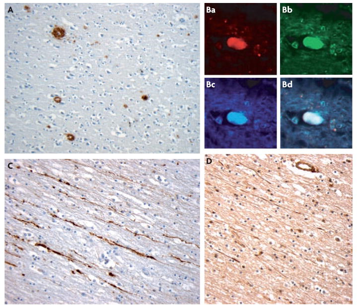
A | representative amyloid-β (Aβ) plaques (brown) found acute following a single incidence of traumatic brain injury (TBI) caused by a fall in an 18 year old male. The survival time from injury was just 10 hours. Plaques were identified using an antibody specific for Aβ. B | representative immunohistochemistry showing amyloid precursor protein (APP) (Ba), β-site-APP-cleaving enzyme (BACE) (Bb) and presenilin-1(PS-1) (Bc) co-accumulating (Bd) in the disconnected terminal of an axon, known as an axon bulb. C | Demonstration of axonal pathology using APP immunohistochemistry. APP (brown) accumulates within the tortuous varicosities along the length of damaged axons. D | Increased neprilysin immunoreactivity (brown) is also observed in damaged axons following TBI. Panel B is reproduced, with permission, from REF. 42 © (2009) International society of Neuropathology.